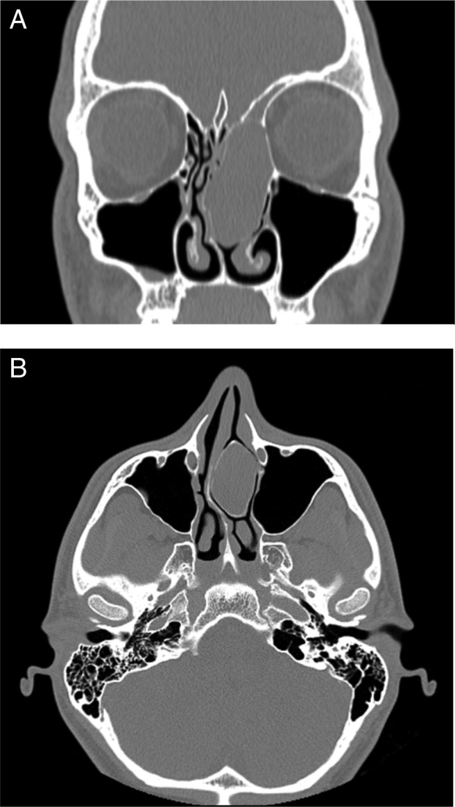
Figure 1.

Computed tomography (C–) of patient 1. (A) Coronal. A 4.2 × 3.5 × 2.2-cm nasal mass extends from the left frontal sinus through the anterior ethmoid until the inferior turbinate; the left frontal sinusitis is also seen. (B) Axial. Mass that fully occupies the nasal cavity, causing right septal deviation.
