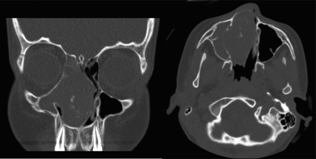
Figure 6.
(A) Coronal view. (B) Axial view of a computed tomography of the sinus in patient 4, which depicts a massive 5.2 × 5.5 × 3.5-cm well-defined bony mass with cortical remodeling, extending to the frontoethmoidal regions. A severe mass effect is seen: there is lateral bowing of the right maxillary sinus, left septal deviation, and elevation of the right fovea ethmoidalis. There was no significant enhancement with contrast. All right-sided sinuses were opacified.

