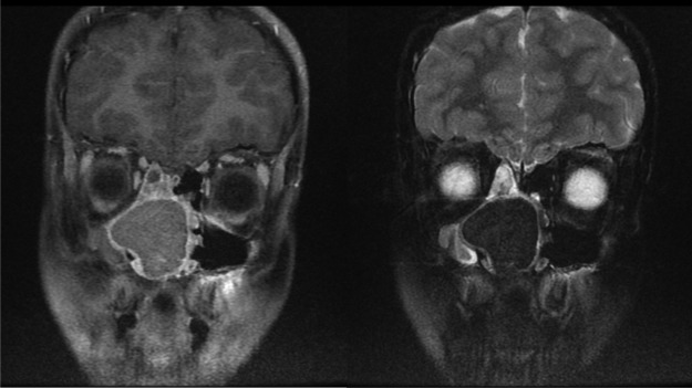
Figure 7.
Patient 4. Magnetic resonance image, (A) T1, (B) T2, showing a large expansile lesion in the right nasal cavity (5.4 × 6.1 × 3.6 cm) extending into the frontoethmoid recess, remodeling of the medial wall of the right maxillary sinus with lateral bowing, and severe nasal septum deviation. There was a T1 hyperintensity and T2 hypointense signal intensity without enhancement and a rim of high T2 signal associated with linear enhancement. Right frontal, ethmoid, and maxillary sinuses are opacified, the right maxillary with inspissated secretions.

