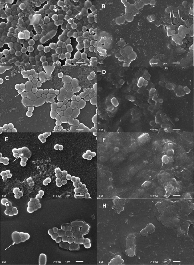
FIG 3.

Scanning electron micrographs of S. aureus dual-species biofilms. Images correspond to 5-h biofilms formed by S. aureus IPLA16 with E. faecium MMRA (A and B), L. plantarum 55-1 (C and D), L. pentosus A1 (E and F), or L. pentosus B1 (G and H) following a 4-h treatment with SM buffer (left) or phiIPLA-RODI (right). White arrows indicate cells from the accompanying species.
