
FIG 4.
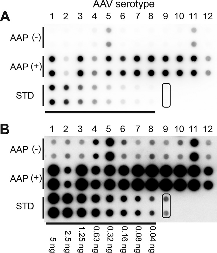
Quantitative dot blot assay to determine titers of VP3 capsids produced in the presence or absence of AAP. (A) AAP-independent assembly of VP3 proteins of AAV1 to -12 assessed by a quantitative dot blot assay in an experiment performed in biological duplicates. VP3-only particles derived from AAV1 to -12 that contained a double-stranded AAV-CMV-GFP genome were produced in 6-well plates in the presence or absence of their cognate AAPs. Benzonase-resistant DNA was recovered from 7% of the samples obtained from each well, blotted onto a nylon membrane together with standards (STD) (i.e., linearized pEMBL-CMV-GFP plasmid), and probed with a 32P-labeled GFP probe. Pairs of dots in each combination represent the results obtained from two separate transfections. The pair of dots indicated with a rounded rectangle are negative controls. (B) Same blot as the one shown in panel A. The signals were intensified equally across the entire image by using ImageJ.