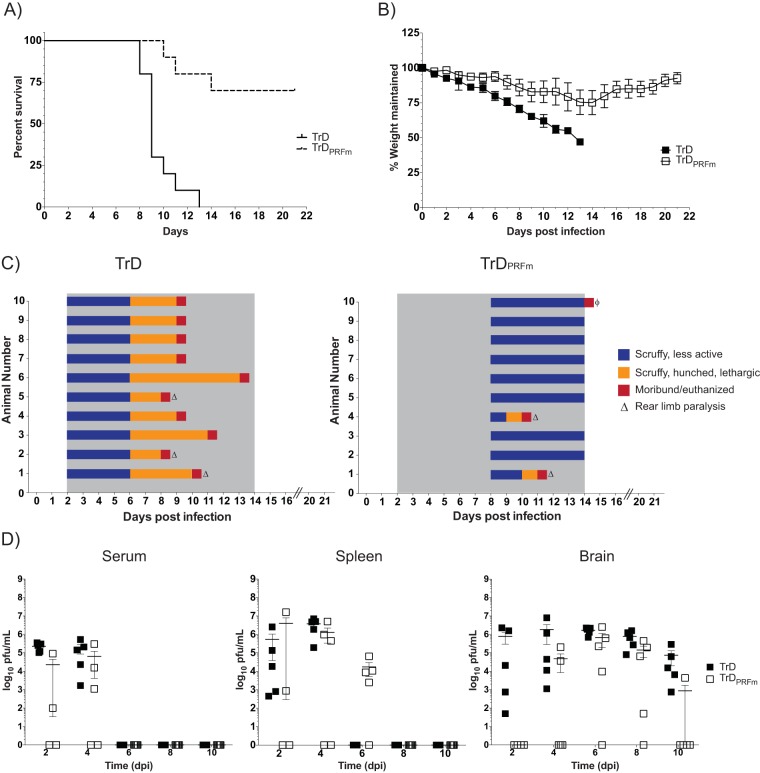
FIG 4.
Ablation of −1 PRF strongly attenuates VEEV pathogenesis. (A) BALB/c mice were infected with VEEV TrD or VEEV TrDPRFm by aerosol exposure. Animals were monitored for 21 days postchallenge, and survival curves were determined. The data plotted represent those for 10 animals per group. (B) Mice were monitored for weight loss daily over 21 days. The percentage of weight maintained (relative to the starting weight) was determined. The data plotted represent the mean values and standard deviations for 10 animals per group. (C) Mice were also monitored at least daily for clinical symptoms of disease over 21 days. Data are plotted per animal per day. The gray shaded area indicates the time frame when clinical disease was observed in VEEV TrD-infected mice. ϕ, one animal had to be euthanized due to self-mutilation. Necropsy indicated no signs of disease in this mouse. (D) Mice were infected as described in the legend to panel A and were sacrificed at 2, 4, 6, 8, and 10 dpi. Brain, spleen, and serum were harvested. Viral titers were determined by plaque assays. The data plotted represent means and standard errors of the means for five animals per condition. Filled and open squares, VEEV TrD and VEEV TrDPRFm, respectively. Samples without detectable plaques were plotted as 1 PFU/ml.

