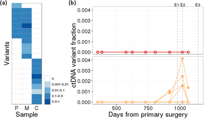
Figure 7. Detection of ctDNA variants is highly specific to tumour origin.
(a) Variant frequency of 20 somatic tumour variants identified by targeted capture sequencing (upper 11 specific to the original CRC primary [P] and liver metastasis [M]; lower 9 specific to second cholangiocarcinoma [C]) as assayed by a patient-specific multiplex amplicon assay in tumour tissue DNA. (b) ctDNA variant fraction over multiple timepoints post-primary surgery. The 11 CRC primary/metastasis variants (upper panel, red) and 9 cholangiocarcinoma variants (lower, orange) are plotted separately. Each line refers to a separate variant. E1: detection of liver lesion. E2: surgery to remove second primary. E3: clinical detection of a lung metastasis by CT scan.

