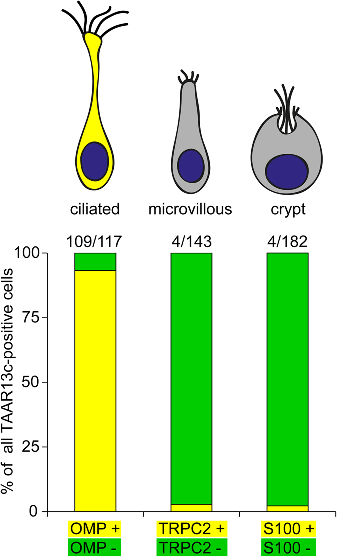
Figure 2. Quantitative evaluation of co-localization of TAAR13c-positive neurons with OSN cell type markers.

Labeling was performed as described in Fig. 1, and over one hundred TAAR13c-labelled cells were evaluated for co-localization with several cell type markers. Results are shown as bar graph below the schematic representation of the three cell types examined. Co-localization is indicated by yellow, non-co-localization by green segments. Nearly all TAAR13c-positive cells were also positive for OMP, a ciliated neuron marker. In contrast, almost no co-localization with TAAR13c staining was observed for TRPC2 and S100, markers for microvillous and crypt neurons, respectively.
