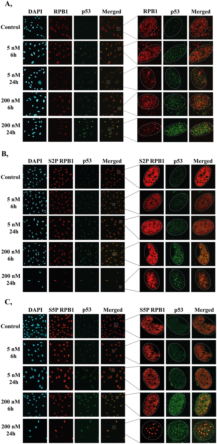
Figure 2. p53 and RPB1 co-localise to discrete nuclear foci in U2OS cells upon ActD-induced transcription elongation blockage.
(A–C) Co-immunostaining with p53 (green) and RPB1 (red), S2P RPB1 (red) or S5P RPB1 (red), respectively. Only chromatin-bound proteins are detected, because nuclear soluble proteins were eliminated during the immunostaining process (see Methods). Immunostainings were performed both under normal conditions and after 6 and 24 h treatments of 5 and 200 nM doses of ActD. Staining with DAPI (blue) was used to visualise nuclei.

