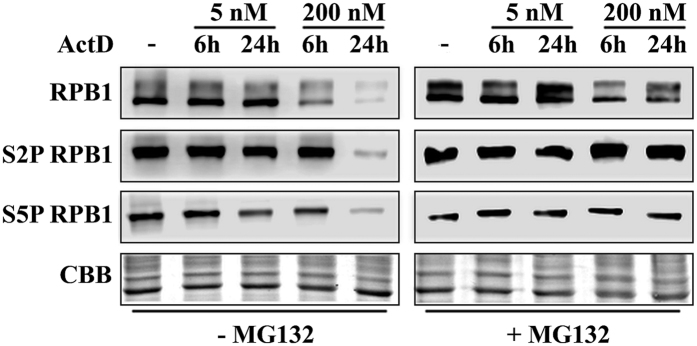
Figure 4. ActD treatment destabilises RPB1 through proteasome-mediated degradation.
Western blot detection of RPB1, S5P RPB1 and S2P RPB1 proteins in U2OS cells both under normal conditions and when treated with 5 and 200 nM ActD. Each experiment was performed in the absence (left panel) or in the presence of 20 μM MG132 proteasome inhibitor (right panel). Coomassie Brilliant Blue staining was used to show the equal loading of the samples.

