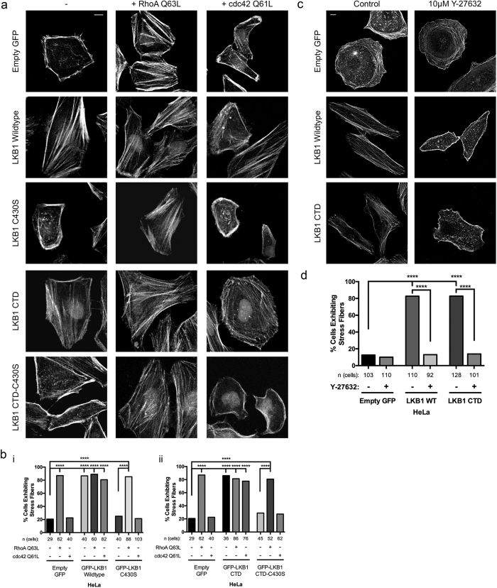
Figure 2. LKB1 signals via the RhoA-ROCK pathway to promote stress fiber assembly.
(a) Immunofluorescent images of HeLa cells expressing GFP-LKB1 alone (−) or while co-expressing constitutively active RhoA (Q63L) or constitutively active cdc42 (Q61L) and stained with phalloidin-555. (b) Percent of cells containing lateral stress fibers was quantified 24 hours after transfection. (i) Percentage of cells containing stress fibers for empty GFP control, GFP-LKB1 Wildtype, and GFP-LKB1 C430S with their respective Rho-GTPase mutants. (ii) Percentage of cells containing stress fibers for empty GFP control, GFP-LKB1 CTD, and GFP-LKB1 CTD-C430S with their respective Rho-GTPase mutants. (c) Immunofluorescent images of HeLa cells expressing GFP-LKB1 with or without 10 μM Y-27632 ROCK inhibitor treatment and stained with phalloidin-555. (d) Percentage of cells containing stress fibers in response to ROCK inhibitor treatment. Significance was measured between comparisons using a 2-tailed Chi-squared analysis with a p-value of 0.05, where ****p ≤ 0.0001. Scale bar: 10 μm.

