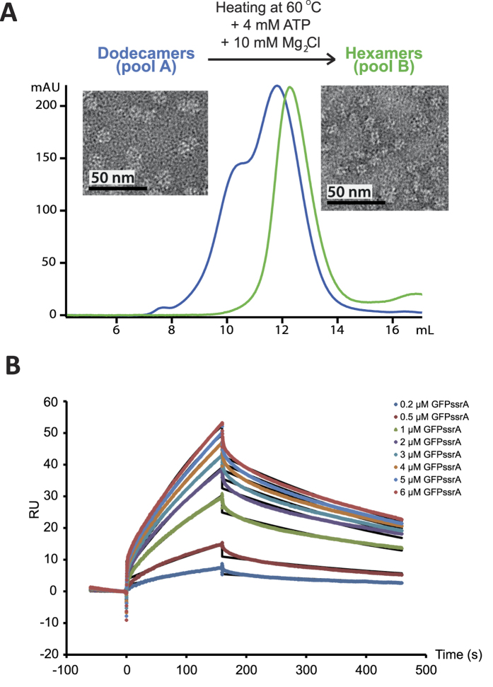
Figure 1. Purification and biophysical characterization of the MjPAN complex.
(A) Superose 6 column chromatography purification profiles of the dodecameric/hexameric mixture (blue chromatogram) and the hexameric (green chromatogram) forms of MjPAN complexes. Negative stain transmission electron micrographs of the MjPAN complex in its dodecameric (pool A) and hexameric (pool B) forms are shown next to the corresponding chromatogram. (B) Kinetic analysis of the interactions of PAN with GFPssrA. The colored lines represent binding responses for injections of protein analyte at specified concentrations (μM) over the PAN-coated surface. The kinetic data were fitted (black curves) by a 1:1 Langmuir binding model45 which describes monovalent analyte binding to a single site on the immobilized ligand (see SI Material and Methods).

