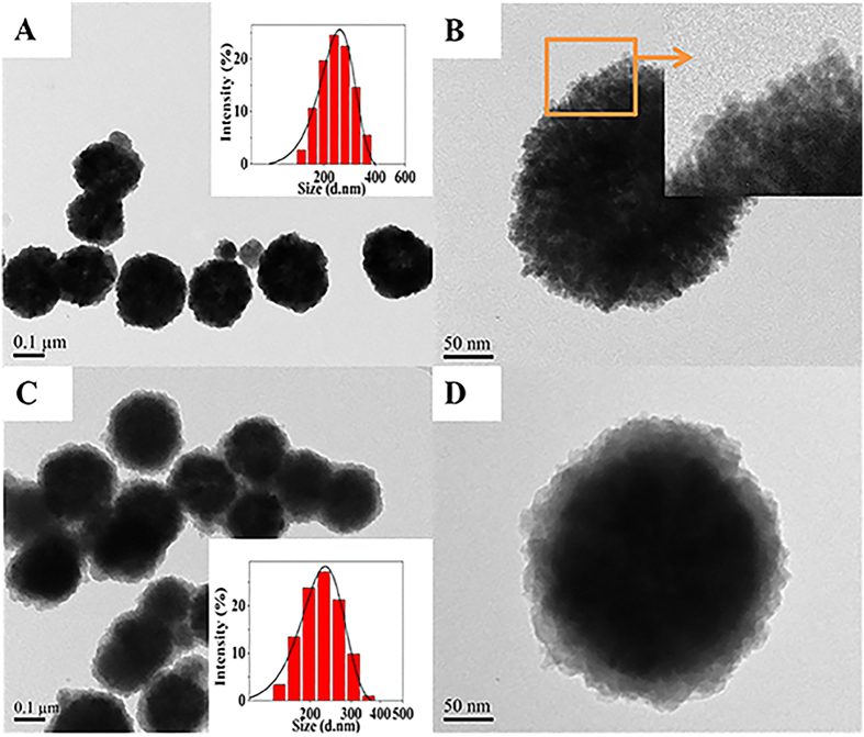
Figure 1. Morphology of CoFe2O4 and CoFe2O4@MIL-100(Fe).
TEM images of (A,B) CoFe2O4 and (C,D) CoFe2O4@MIL-100(Fe); inset: size distributions from dynamic light scattering. The TEM images indicate that both CoFe2O4 and CoFe2O4@MIL-100(Fe) MNPs exhibited excellent nanoscale and mesoporous properties.

