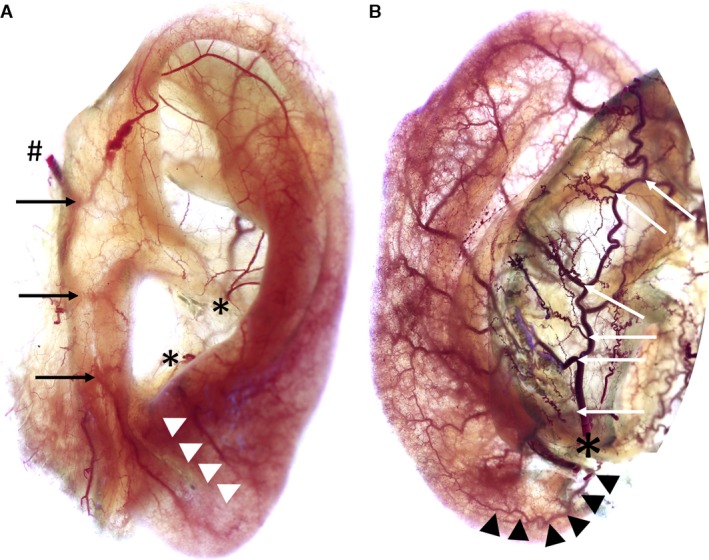
Figure 2.

Images show anterior (A) and posterior (B) view of left auricles stained according to ‘Spalteholz’ method. Image A: #Superficial temporal artery, black arrows mark superior/middle/inferior anterior auricular arteries, *Helical root and antitragal perforator, white arrowheads mark branch of antitragal perforator supplying the earlobe. Image B: *Posterior auricular artery, white arrows indicate the perforating and non‐perforating branches, black arrowheads mark the inferior anterior auricular artery running towards the earlobe.
