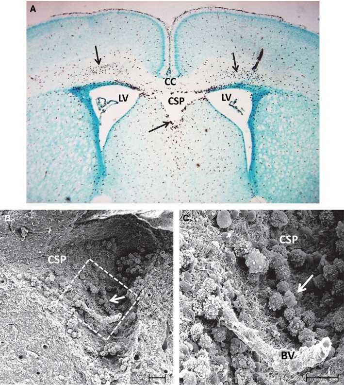
Figure 1.

(A) A coronal section of the brain in a 3‐day‐old rat showing the cavum septum pellucidum (CSP) located in between the lateral ventricles (LV) beneath the corpus callosum (CC). A large number of amoeboid microglia (arrows) stained with the antibody OX 42, a specific marker for microglia, can be seen in the CC and the CSP. (B) A scanning electron micrograph of the CSP in a 3‐day‐old rat. The squared area in (B) is shown at higher magnification in (C). A blood vessel (BV) appears to course through the CSP lumen. Note the occurrence of a large number of amoeboid microglia (arrows) depicted in (B and C), which are adherent to the walls of CSP made up of a felt‐work of nerve and glial fibres. Scale bars: 40 μm (B); 20 μm (C).
