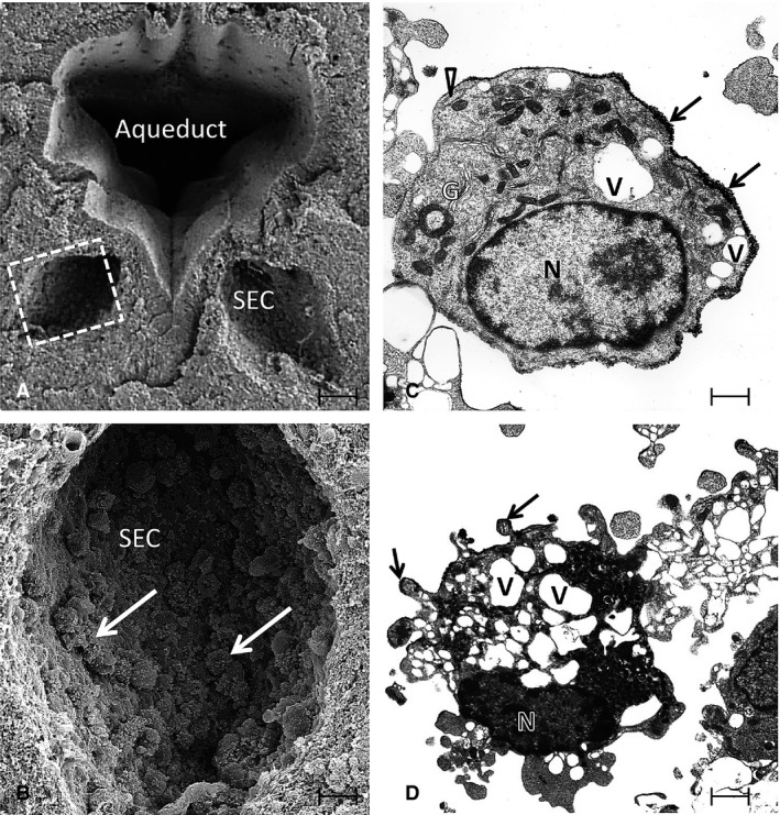
Figure 2.

(A) A scanning electron micrograph (SEM) of the aqueduct in the brain of a 3‐day‐old rat. Note the two symmetrical subependymal cysts (SEC), one on each side. The squared area in one of the SECs in (A) is shown at higher magnification in (B). A number of amoeboid microglia (arrows) are seen adhering to the walls of the SEC. (C and D) Transmission electron microscopic images of the amoeboid microglia from re‐embedded material used for SEM. The amoeboid microglial cell in (C) has a smooth cell surface and shows a nucleus (N), well‐developed Golgi apparatus (G), cisternae of rough endoplasmic reticulum (arrowhead) and many vacuoles (V). The cell in (D) shows a nucleus (N), vacuoles (V) and many blebs at the surface. Arrows in (C and D) indicate sputter‐coated gold on the cell surface. Scale bars: 70 μm (A); 20 μm (B); 1.4 μm (C); 0.8 μm (D).
