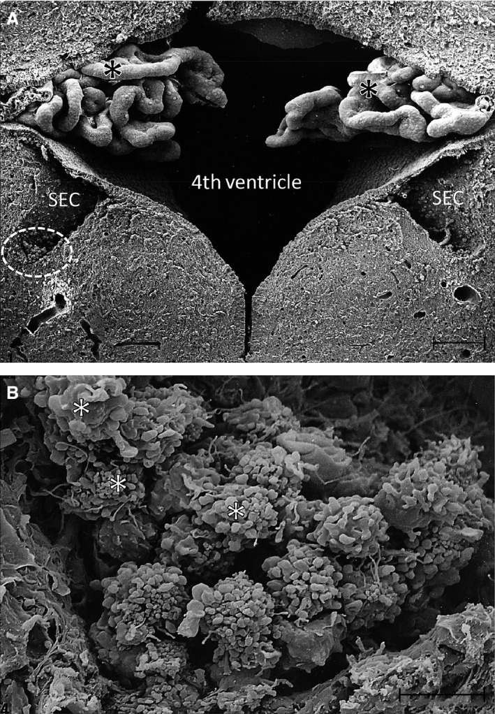
Figure 3.

(A) A scanning electron micrograph of the fourth ventricle in the brain of a 3‐day‐old rat with a subependymal cyst (SEC) on each side. Note the convoluted choroid plexus (asterisks) in the ventricle. The circled area in (A) indicates a portion of one of the SECs shown at a higher magnification in (B). A prominent cluster of amoeboid microglia is seen in the SEC in (B). The cells (asterisks) show prominent blebs at the surface with some of them showing long filopodia. Scale bars: 100 μm (A); 10 μm (B).
