
Figure 3.
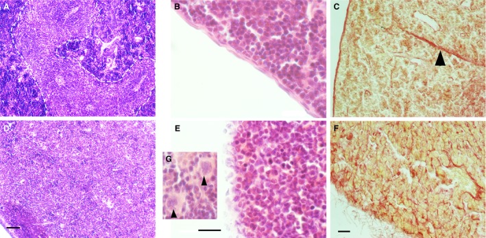
A morphologically distinct white pulp fails to develop and the spleen begins to degenerate in spinal muscular atrophy (SMA) mice. H&E staining of late‐symptomatic (P8) spleens. (A) In the control spleen, white pulp (dotted line) is clearly separated from red pulp. (D) In the SMA spleen, red and white pulp are undiscernible from one another, and there is an accumulation of large cells with lobulated nuclei (arrowhead, G). Scale bar: 50 μm. (B) Control spleen displays an intact splenic capsule, whereas the SMA spleen lacks a smooth and organised capsule‐like structure, instead presenting apparent dissociation of capsular cells (E). Scale bar: 20 μm. (C, F) Picro Sirius Red staining of the collagen component of the splenic capsule and trabeculae (arrowhead) in the control and late‐symptomatic (P8) spleen. The splenic capsule and trabeculae are intact in the control spleen (C); however, the SMA spleen lacks trabeculae and shows dissociation of the capsule (F). Scale bar: 50 μm.