
Figure 1.
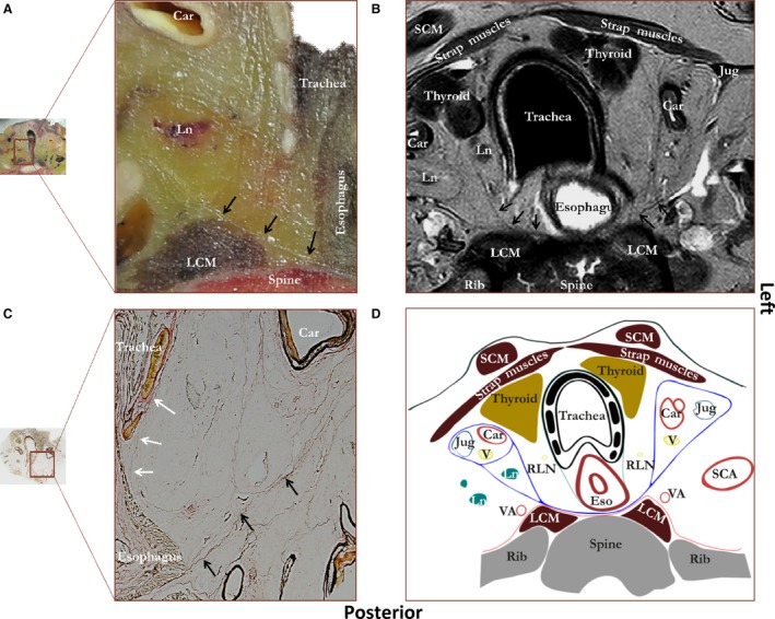
Figure showing a photograph of an unprocessed transverse tissue section at the level of the thyroid gland (A) with a corresponding MRI image (B), microscopic tissue section (C) and schematic drawing (D). Staining was performed according to Verhoef‐von Gieson, which stains elastin black‐blue and collagen light red‐pink. Black arrows, alar fascia; white arrows, thin visceral fascia enveloping esophagus and trachea. Car, carotid artery; Eso, esophagus; Jug, internal jugular vein; LCM, longus colli muscle; Ln, lymph node; Rln, recurrent laryngeal nerve; SCA, subclavian artery; SCM, sternocleidomastoid muscle; V, vagus nerve; VA, vertebral artery. Blue line, alar fascia and carotid sheaths; green line, visceral fascia; red line, perivertebral fascia.