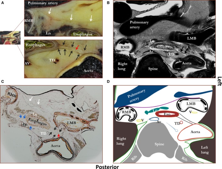
Figure 2.

Figure showing a photograph of an unprocessed transverse tissue section at the level of the tracheal bifurcation (A) with a corresponding MRI image (B), microscopic tissue section (C), and schematic drawing (D). Staining was performed according to Verhoef‐von Gieson, which stains elastin black‐blue and collagen light red‐pink. Black arrows, double layer of connective tissue between esophagus and aorta (proposed name ‘aorto‐esophageal ligament’); blue arrows, a connective tissue layer coursing from aorta to right pleural reflection (proposed name ‘aorto‐pleural ligament’); white arrows, a layer of connective tissue coursing from right to left main bronchus; red arrow, a blood vessel. Av, azygos vein; LMB, left main bronchus; Ln, lymph node; RMB, right main bronchus; TD, thoracic duct. Green line, pleura; purple line, connective tissue layer coursing from left to right main bronchus; black line, connective tissue layer coursing from aorta to esophagus; gray line, connective tissue layer coursing to the right pleural reflection.
