
Figure 1.
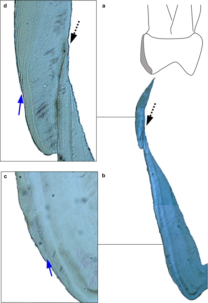
Hypoplastic defect and Retzius periodicity (zoom in to see Retzius lines). (a) Human deciduous maxillary second molar mesio‐lingual enamel highlighted in grey. (b) The same region imaged using a polarizing lens. Dashed arrow points to a hypoplastic defect associated with an accentuated marking. Magnification = 4×. (c) Blue arrow points to Retzius lines that formed before the hypoplastic defect. Magnification = 20×. (d) Blue arrow points to Retzius lines that formed after the hypoplastic defect. The stress event did not prevent secretory ameloblasts from recovering, as these cells had a functional Tomes process (separate rods are visible) that deposit enamel at a slightly accelerated rate.