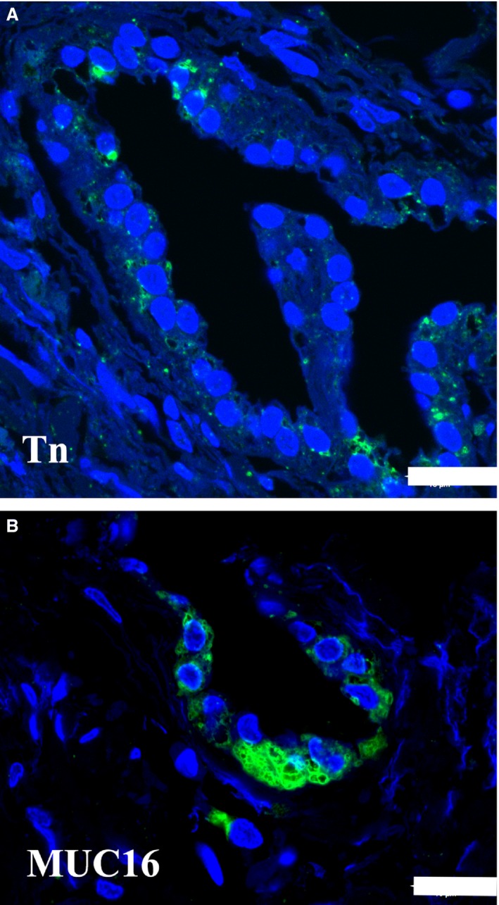
Figure 4.

Immunohistochemical demonstration of the Tn‐antigen and MUC16 in the human endolymphatic sac. The green fluorescence marks the detection of the mucin. The blue fluorescence is caused by 4,6‐diamino‐2‐phenylindole‐2HCl stain in the nuclei. (A) There is a scattered reaction with anti‐Tn in a few saccular epithelial cells. Scale bar: 20 μm. (B) An intense labeling of the MUC16 antibody is present mostly at the basal part of the epithelial cells. Scale bar: 20 μm.
