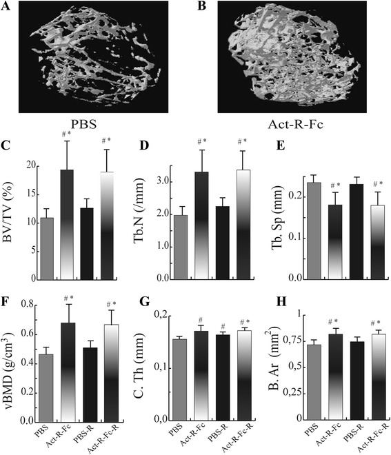
Fig. 1.

Reconstructed 3D images of the distal femur of control (a) and Act-R-Fc treated mice (b) showing a significant increase in bone mass in the treated group. μCT analysis of the distal femur showing (c) increased bone volume (BV/TV), (d) trabecular numbers (Tb.N), (e) decreased trabecular separation and (f) increased volumetric bone mineral density (vBMD) in ActRIIB-Fc-treated mice compared to controls. Significant increases in (g) cortical thickness and (h) mean total cross-area parameters were also noted. Running combined with ActRIIB-Fc treatment did not further improve these parameters N = 7-9 per group. # = p < 0.05 compared to PBS, * = p < 0.05 compared to PBS-R
