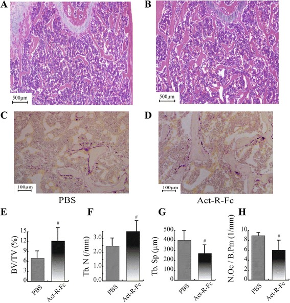
Fig. 3.

Histological images (H&E staining) of the distal femur showing an increase of trabecular numbers in ActRIIB-Fc treated group (b) compared to PBS controls (a). TRACP staining of distal femurs of control (c) and Act-R-Fc treated mice (d). Histomorphometric analysis showed increased bone volume (e) and trabecular number (f) and decreased trabecular separation (g) as in μCT. Interestingly, the number of TRACP positive osteoclasts was reduced in Act-R-Fc-treated mice (h). The region of interest from which the trabecular islands and the stained osteoclasts were analyzed consisted of an 800μm x 1200μm area near the distal growth plate. N = 8-9 per group. # = p < 0.05 compared to PBS
