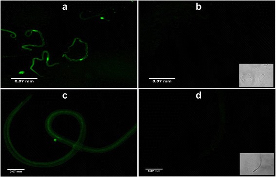
Fig. 3.

Successful uptake of siRNAs by B. malayi microfilariae and infective larvae. The images show successful siRNA uptake by B. malayi microfilariae released by siRNA-treated female parasites of different groups (a), unexposed control microfilariae (with phase contrast image at right bottom) (b), siRNA-treated infective larvae (c), and unexposed control infective larvae (with phase contrast image at right bottom) (d) after 24 h under the fluorescent microscope. The images were taken under FLoid Cell Imaging Station (Life technologies, US) at 520 nm emission
