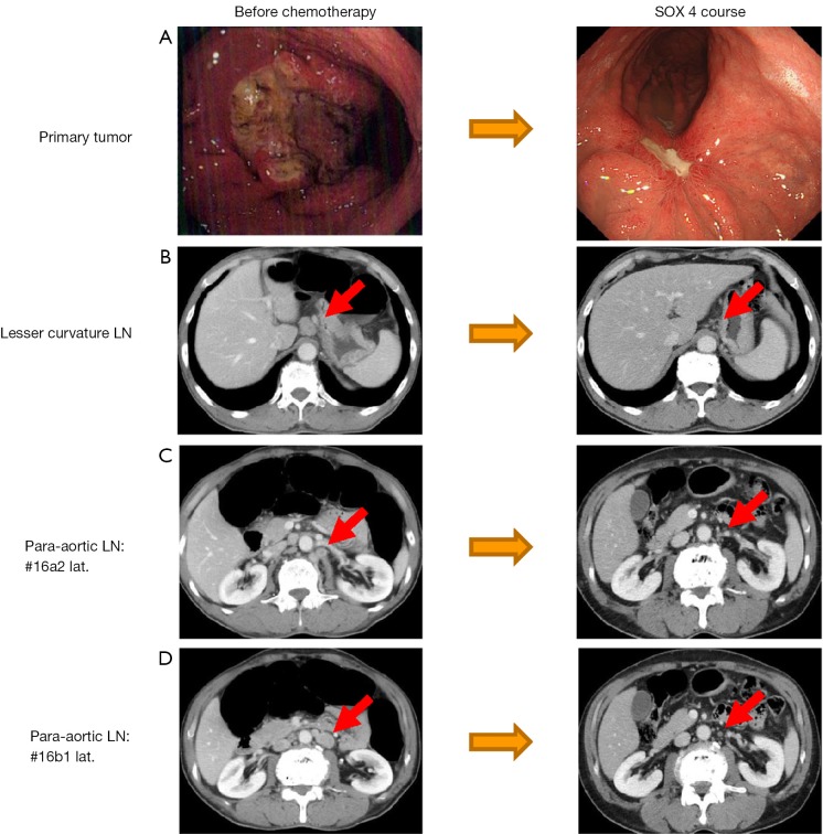
Figure 2.
The case presentation in category 1. Endoscopic images (A) show the primary tumor at the initial diagnosis and after 4 cycles of SOX therapy. Shrinkage of the primary tumor was obtained and part of the ulcer was scarred; CT images (B-D) show metastatic LNs at the lesser curvature and the para-aortic region. Similarly, shrinkage of the LNs was obtained, the response was considered to be a PR (76.9%), which was confirmed by the RECIST criteria.

