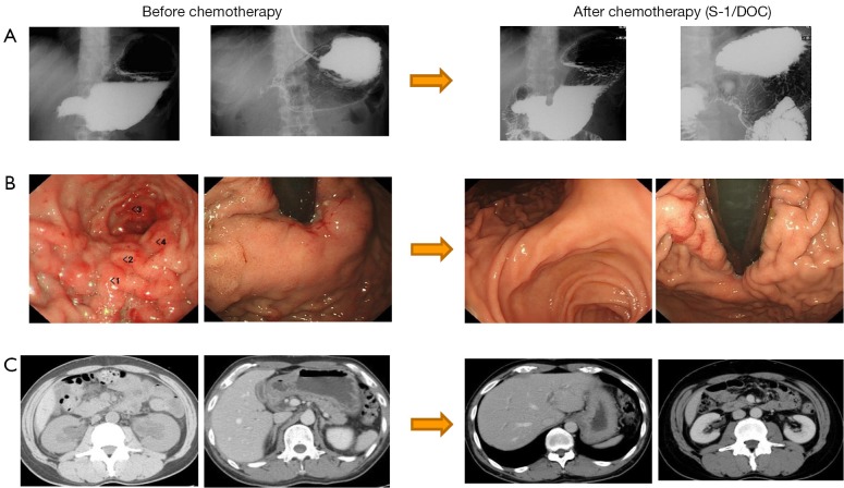
Figure 4.
The case presentation in category 3. An upper gastrointestinal image (A) and an endoscopic image (B) show pyloric stenosis and the thickening of the gastric wall; a CT image (C) shows bilateral hydronephrosis. After 6 cycles of S-1/DOC therapy, the patient completely recovered from hydronephrosis and oral intake became possible.

