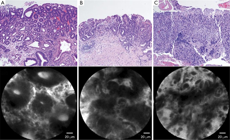
Figure 1.
Features of confocal endomicroscopy. (A) Dysplasia, dark epithelium with irregular and varying thickness is observed; (B) differentiated adenocarcinoma, disorganized epithelium with dark and irregular glands is observed; (C) undifferentiated adenocarcinoma, dark and irregular cells with no identifiable glandular structures are observed. (H&E, ×100).

