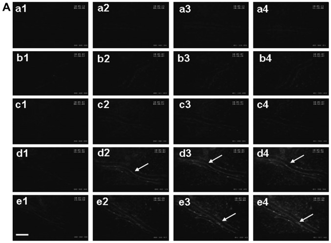
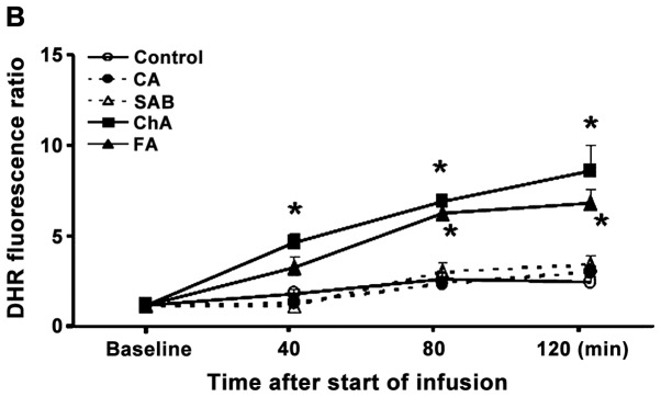
Figure 2.
Effect of phenolic acids on DHR fluorescence intensity in rat mesenteric venular wall. (A) Representative images of the changes in DHR fluorescence intensity of the H2O2-sensitive probe DHR in the mesenteric venular wall in the (a) control, (b) CA, (c) SAB, (d) CA and (e) FA groups at (1) baseline, (2) 40, (3) 80 and (4) 120 min, respectively. The arrow indicates DHR fluorescence on the venular wall; scale bar, 50 µm. (B) Time course of changes in the ratio of DHR fluorescence on the venular walls in different groups. Data are presented as means ± SE of six animals. *P<0.05 vs. control group. DHR, dihydrorhodamine 123; CA, caffeic acid; SAB, salvianolic acid B; FA, ferulic acid.


