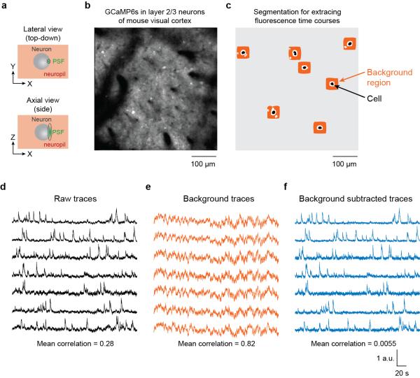
Figure 3.
Subtracting neuropil contamination from raw fluorescence time courses. (a) In two-photon imaging, the point spread function (PSF) is elongated in the axial dimension even in high numerical aperture systems. Pixels within the borders of cell bodies still contain signals from the surrounding neuropil. (b) The GECI GCaMP6s was expressed in mouse visual cortex neurons, resulting in brightly labeled cell bodies and neuropil. (c) Binary masks for cell body ROIs (black) were identified semi-automatically and neuropil regions were algorithmically constructed (avoiding pixels belonging to other potential cell bodies or black regions). (d) Raw traces for the fluorescence time courses of the selected cells. (e) Fluorescence time courses for the background regions for each selected cell. Note the high temporal correlation. (f) Fluorescence time courses after background subtraction. Note the reduced mean correlation141. All traces have been scaled to the same maximum height to better exhibit details in the time courses. Figure credit: J. N. Stirman, Y. Yu, S. L. Smith

