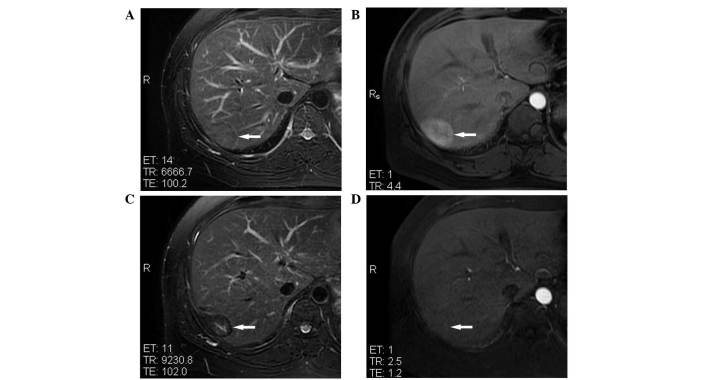
Figure 1.
MRI of an inflammatory pseudotumor of the liver, with a maximal diameter of 45 mm, in hepatic segment VII of a 38-year-old male patient prior to and following microwave ablation. (A) T2WI showing inflammatory pseudotumor of the liver isointensity (white arrow) prior to treatment. (B) Contrast-enhanced MRI showing IPL hyperintensity (white arrow) in the arterial phase prior to treatment. (C) T2WI showing ablated lesion hypointensity (white arrow) at 1 year post-treatment. (D) Contrast-enhanced MRI showing the lack of enhancement of the ablated lesion (white arrow) in the arterial phase at 1 year post-treatment. MRI, magnetic resonance imaging; WI, weighted imaging.

