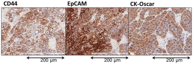
Figure 6.
Immunohistochemical evaluation of the resected primary tumor. CK-Oscar staining was used a positive control to indicate tumorous tissue. In advanced GC, almost all cancer cells were stained with EpCAM and CD44. By contrast, in early GC, EpCAM-stained cells were present in almost all cancer cells, but only a few CD44-stained cells were present (original magnification, ×400). GC, gastric cancer; EpCAM, epithelial cell adhesion molecule; CD44, cluster of differentiation 44.

