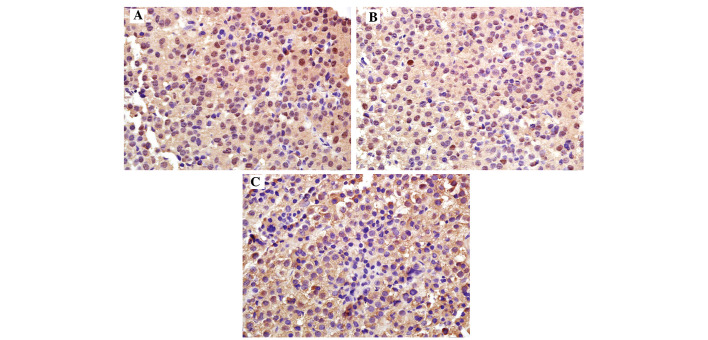
Figure 2.
PTEN expression in normal pituitary tissue and pituitary adenomas (magnification, ×200). PTEN was mainly expressed in the cytoplasm, displaying as a brownish-yellow stain. The expression of PTEN in (A) normal pituitary tissues was higher compared with that in (B) the non-invasive group (P<0.05), which was in turn higher compared with that in (C) the invasive group. The positive rate had a significantly decreasing trend with the increasing degree of tumor invasiveness. PTEN, phosphatase and tensin homolog deleted on chromosome 10.

