
Figure 3.
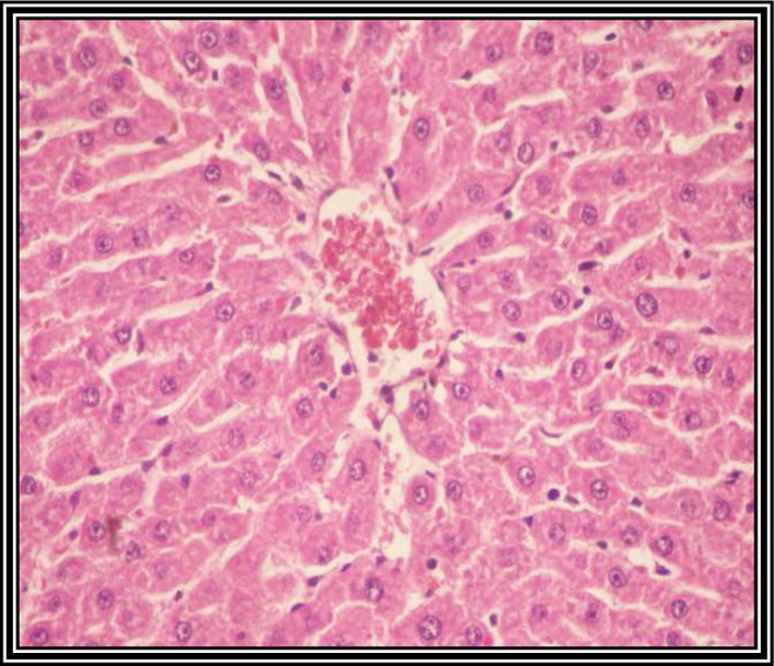
Photomicrograph of a liver section from untreated control rats showing normal organized hepatic tissue with normal hepatocytes (hematoxylin and eosin staining; magnification, ×400).
Official websites use .gov
A
.gov website belongs to an official
government organization in the United States.
Secure .gov websites use HTTPS
A lock (
) or https:// means you've safely
connected to the .gov website. Share sensitive
information only on official, secure websites.
Photomicrograph of a liver section from untreated control rats showing normal organized hepatic tissue with normal hepatocytes (hematoxylin and eosin staining; magnification, ×400).