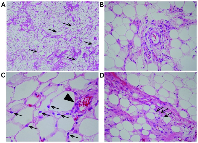
Figure 2.
Hematoxylin and eosin-stained sections of lipomatous angiomyofibroblastoma. (A) Low-power magnification (x40) showing numerous fat cells, together with medium-sized vessels and pseudoangiomatous spaces (arrows). (B) Spindle and rounded tumor cells proliferating singly or in clusters in perivascular fibrotic areas (magnification, ×400). (C) Spindle or epithelioid neoplastic cells (arrows) scattered singly between fat cells and in a nest-like pattern around a vessel (arrowhead) (magnification, ×600). (D) A few vacuolated cells (arrows) were identified near the tumor and fat cells (magnification, ×400).

