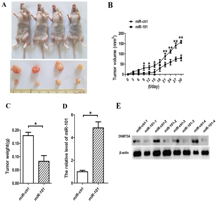
Figure 6.
miR-101 inhibits lung cancer progression in vivo. (A) Small animal imaging analysis was used to assess the tumor volume in situ during the fourth week of tumor development. The flanks of four nude mice were injected with lentiviral vector (LV)-miR-101-transfected cells (left flank) and LV control (right flank), respectively. The lower portion reveals the morphology of mice injected with miR-101 and miR-ctrl. (B) Tumor growth curves. (C) Tumor weight. (D) Expression levels of miR-101 were measured by reverse transcription-quantitative polymerase chain reaction analysis in the tumor tissues from the animals. (E) Expression levels of DNA methyltransferase 3A (DNMT3A) were assessed by western blot analysis in tissues from the animals. β-actin was used as a housekeeping control (*P<0.05, **P<0.01, Student's t-test).

