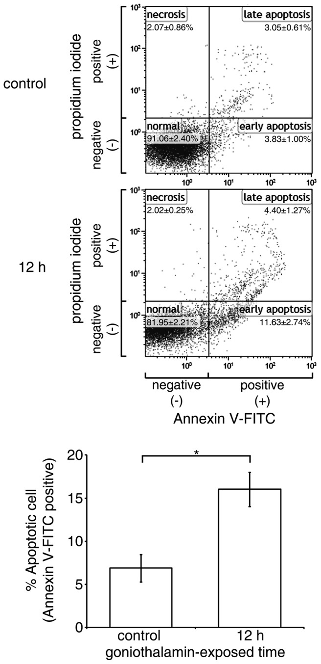
Figure 4.

Increases of cell surface phosphatidyl-serine presentation in HeLa cells by goniothalamin. The Cell surface presentation of phosphatidyl-serine presentation on HeLa cells treated with 15 µM goniothalamin for 12 h was detected via Annexin V assessment. The percentage in each quadrant indicates the levels of normal cells (annexin V-FITC−/propidium iodide−), early apoptotic cells (annexin V-FITC+/propidium iodide−), late apoptotic cells (annexin V-FITC+/propidium iodide+) and necrotic cells (annexin V-FITC−/propidium iodide+). These results indicated that goniothalamin induced phosphatidyl-serine exposure, indicative of apoptosis induction. The results shown are representative data from three independent experiments. Percentages of apoptotic cells are presented as the mean ± standard deviation from at least three independent experiments. *P<0.05, vs. control. FITC, fluorescein isothiocyanate.
