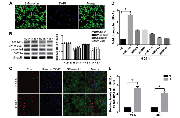
Figure 1.
Effect of anoxia on the general characteristics of rat PASMCs and screening of differentiated miRNA. (A) Identifying PASMCs (x200) with SM-α-actin immunofluorescent staining. (B) Detecting the expression of SM-MHC, SM-α-actin, calponin-1 and SM22α proteins with western blot analysis. Based on a comparison with the control group, *P<0.05. (C) Detecting the proliferative activity of PASMCs with EdU staining. (D) Differential expression of miRNA at 24 h after anoxia. (E) Expression of miR-23a at 24 and 48 h after anoxia. *Compared to the control group, P<0.05. PASMCs, pulmonary artery smooth muscle cells.

