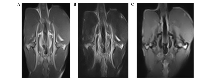
Figure 2.
Hip magnetic resonance coronal scanning images of rabbits with necrosis of the femoral head. (A) Bilateral femoral heads exhibited thin thread-like low signals in the T1-weighted image, and the left femoral head was irregular. (B) T2-weighted imaging also exhibted low signal, (C) whereas high signal in the gradient echo short time inversion recovery image of the femoral head, which exhibited no joint effusion.

