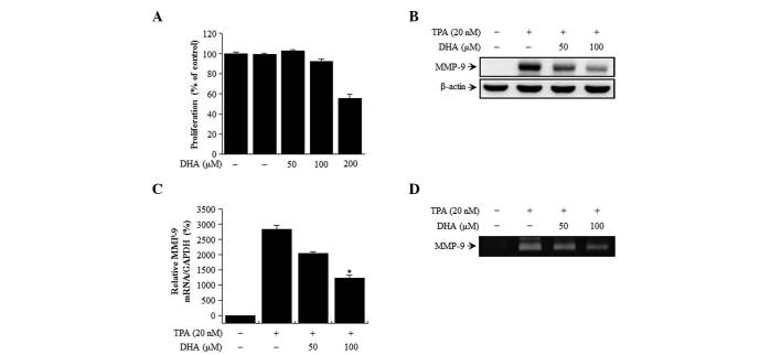
Figure 1.
Effect of DHA on cell viability and TPA-induced MMP-9 expression in MCF-7 cells. (A) The cytotoxicity of DHA was assessed using the MTT assay in cells exposed to the indicated concentrations of DHA for 24 h. The optical density value of control cells was considered as 100%. (B) MMP-9 expression was detected in cell lysates by western blotting, with β-actin used as a loading control. (C) MMP-9 mRNA level was analyzed by reverse transcription-quantitative polymerase chain reaction relative to the level of GAPDH. (D) MMP-9 activity was evaluated in conditioned medium by gelatin zymography. Values represent the mean + standard error of the mean of three independent experiments. *P<0.01 vs. TPA. TPA, 12-O-tetradecanoylphorbol-13-acetate; DHA, docosahexaenoic acid; MMP, matrix metalloproteinase; mRNA, messenger RNA.

