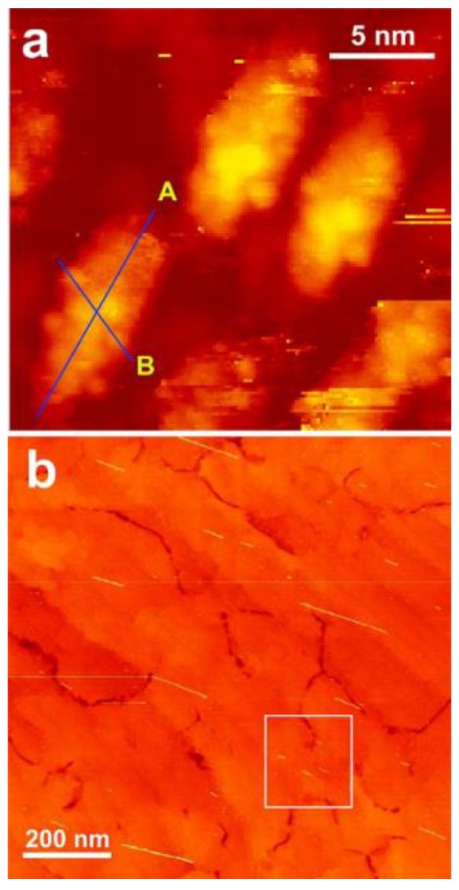
Figure 3.

Scanning tunneling microscopy (STM) images of DNA-CNT hybrids deposited on p-doped Si(110) surface. (a) 21 nm × 21 nm scanning area; (b) 1 μm × 1 μm scanning area. For preparation of the DNA-CNT hybrids, 20 mer DNA (5′-NH2(C-6)GAGAAGAGAGCAGAAGGAGA-3′) was employed. Before the STM imaging, the sample was annealed at 550 °C for 30 min in vacuum (reprinted from reference [52] with permission).
