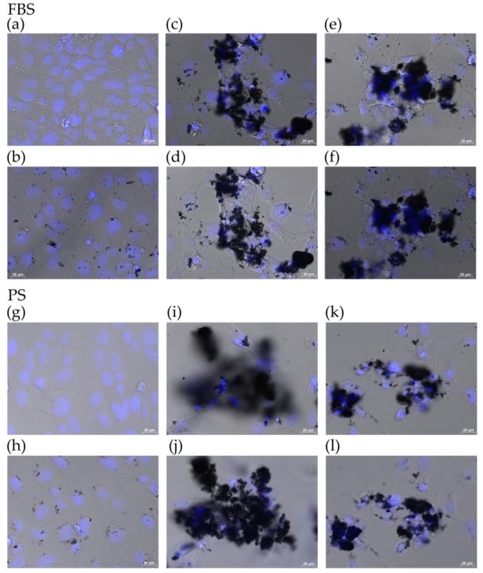
Figure 3.
Live cells imaged by differential interference contrast optics after incubation with bisbenzimide H33342 fluorochrome trihydrochloride for nuclear staining in two dispersants. (a,g) Control; (b,h) PR-1; (c,i) US-1R bottom; (d,j) US-1R top; (e,k) W-220 bottom; and (f,l) W-220 top. “Bottom” indicates that the image was captured when the microscope was focused on the bottom of the culture dish. “Top” indicates that the image was captured when the microscope was focused on cells adhered to the top of agglomerated Flotube 9110.

