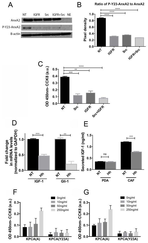
Figure 1. Inhibition of Src and IGF-1R kinases results in decreased phosphorylation of AnxA2 at Tyrosine 23 on the cell surface of human PDAC cells and subsequent decrease in PDAC invasion while stromal factors, IGF-1 and HGF, upstream of IGF-1R and Src kinases, enhance invasion of tumor cells in PDAC cells.
A. Western blot of Panc10.05 human PDAC cells treated with IGF-1R inhibitor (1μM) and Src inhibitor (50 nM) for 60 minutes. AnxA2 was eluted off the surface of PDAC cells with EGTA as previously described (12,26). The levels of total surface AnxA2 and P-Y23-AnxA2 were quantified by Western blot. β-actin was used as a loading control. B. Quantification of relative expression of P-Y23-AnxA2 to total cell surface AnxA2(ratio) C. An invasion assay using PDAC cells treated with IGF-1R and/or Src inhibitors D. qRT-PCR analysis of IGF-1 and Gli-1(control) in hCAFs. The gene expression of IGF-1 and Gli1 was normalized to GAPDH and is shown as a fold change. E. IGF-1 secretion determined by ELISA from single cultures of PDACand hCAF, respectively. F and G. An invasion assay using murine KPCA (A) and KPCA (Y23A) PDAC cells was performed. Serum free media, rIGF-1(C) or rHGF (D) at depicted concentrations were added to the PDAC cell suspension prior to plating. Data are means ± SEM from three technical replicates and representative of at least duplicate experiments. NT-vehicle treatment, Hh-Hh inhibitor (NVP-LDE225 at 1μM) treatment, IGFR-NVP-AEW541 inhibitor, Src-dasatinib inhibitor. ns-not significant, *p<0.05, **p<0.01, ***p<0.001, ****p<0.0001(unpaired student’s t-test).

