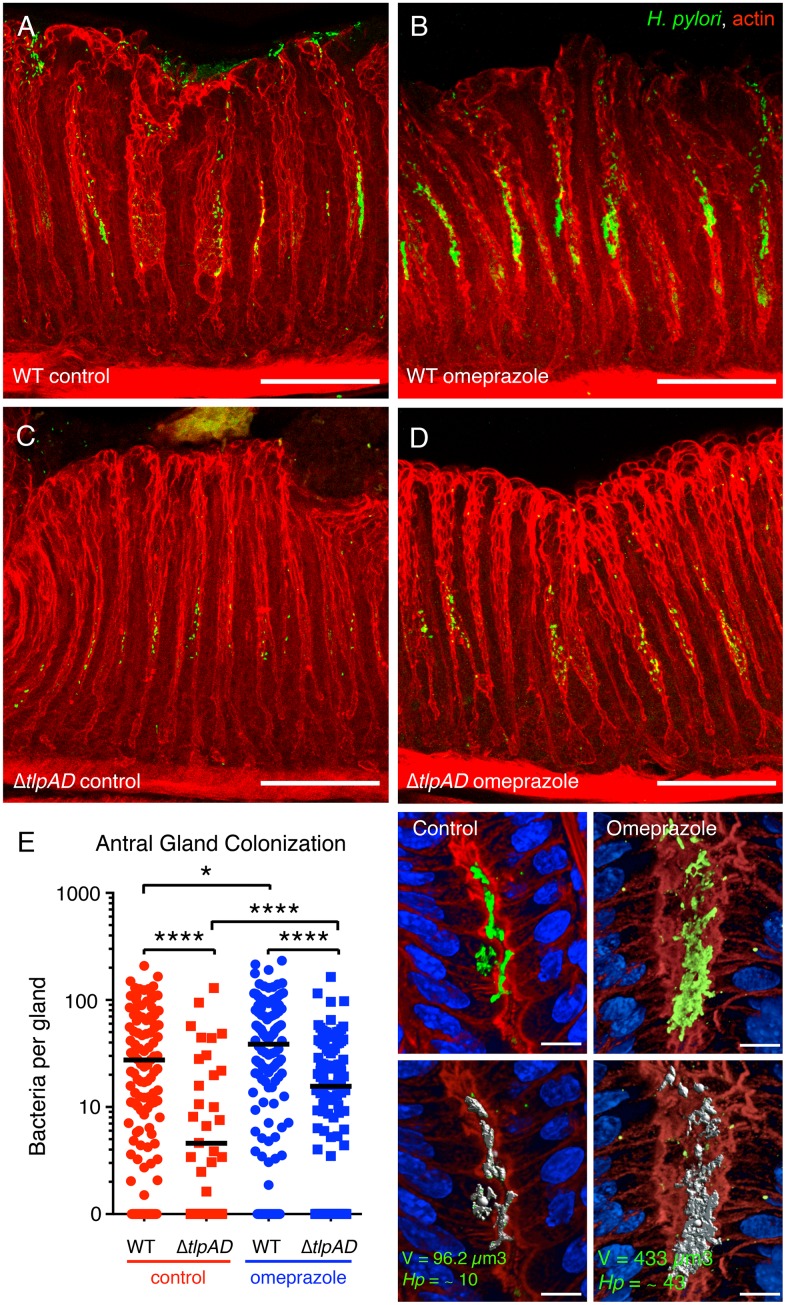
Fig 7. The density of gland-associated H. pylori in the antrum of the stomach is influenced by gastric acidity.
(A-D) 3D confocal immunofluorescence reconstructions of mice infected with wild-type (WT) or ΔtlpAD H. pylori followed by treatment with omeprazole or no treatment. H. pylori are stained in green and actin in red. Scale bar is 100μm for all 4 panels. (E) Quantification of the number of bacteria per gland in the antrum of animals infected with WT or ΔtlpAD treated with omeprazole or untreated. The antral glands of seven animals in each cohort were analyzed for bacterial colonization by 3D volumetric analysis. The number of bacteria in each individual gland is plotted as a scatter point. * P < 0.05, **** P < 0.0001 (Mann Whitney test). The panels on the right show an example of the volumetric measurement of bacteria in gastric glands. 3D confocal reconstructions of glands stained with anti–H pylori (green), phalloidin (red), and DAPI (blue) were generated. Bacterial microcolony signals are identified by fluorescence intensity and size using Volocity software and the identified voxels marked in gray (bottom panels) are measured. The number of bacteria per gland is calculated based on the average voxel volume of 1 bacterium. Scale bar is 10μm for all 4 panels.

