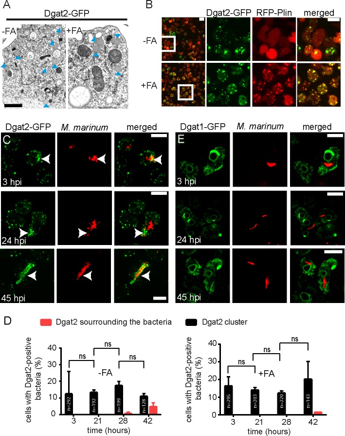
Fig 1. Localization of Dgat1- and Dgat2-GFP during infection of Dictyostelium with M. marinum.
A. LDs with their typical morphology are formed in cells overexpressing Dgat2-GFP even without FA supplementation. Cells that were treated with and without FAs were fixed and processed for EM. Arrowheads label LDs. Scale bar, 1 μm. B.Dynamics of RFP-Plin and Dgat2-GFP in Dictyostelium treated with exogenous FAs. In axenic medium without FA supplementation, RFP-Plin is cytosolic whereas Dgat2-GFP is located on LDs. Upon treatment with exogenous FAs, RFP-Plin translocates to the surface of LDs where it co-localizes with Dgat2-GFP. Dictyostelium expressing both RFP-Plin and Dgat2-GFP was cultured in medium supplemented with FAs and a time-lapse movie was recorded with 5 min frame intervals. Shown is the maximum z-projection of 6 sections spaced 1.5 μm apart. Scale bars, 10 μm. C. Dgat2-GFP-positive LDs cluster at bacterial poles. Dictyostelium expressing Dgat2-GFP was infected with mCherry-expressing mycobacteria. Samples for live imaging were taken at 3, 24 and 45 hpi. Dictyostelium was fed with FA prior to infection. Arrows point to LD clusters. Scale bars, 10 μm. D. Quantification of C. The number of Dictyostelium cells harbouring bacteria that co-localize with LDs aggregates was stable over the time course of infection. Bacteria surrounded by Dgat2-GFP were only observed at late stages, as judged by quantification using z-projections. The statistical significance was calculated with an unpaired t-test (* p<0.05, ** p<0.01). Bars represent the mean and SD of two independent experiments. E. Dgat1-GFP is enriched at the ER and at the perinuclear ER during infection of Dictyostelium with mCherry-expressing M. marinum. Samples for live imaging were taken at 3, 24 and 45 hpi. Dictyostelium was fed with FA prior to infection. Scale bars, 10 μm.

