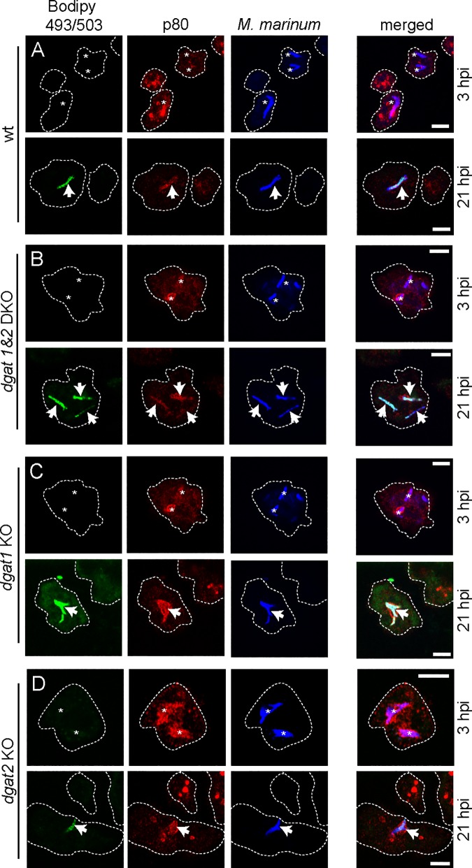
Fig 3. Bacteria accumulate ILIs in the dgat KO mutants.
Cells of (A) wild type, (B) dgat1&2 DKO, (C) dgat1 KO and (D) dgat2 KO and were infected with mCherry-expressing M. marinum. At 3 hpi bacteria are lean (asterisks) whereas at 21 hpi bacteria harbour many ILIs in all cell types (arrows). Cells were fed with FAs prior to infection. At the indicated time points samples were fixed with PFA/picric acid, and MCVs visualized by staining for p80. Bacterial ILIs were stained with Bodipy 493/503. Scale bar, 5 μm.

