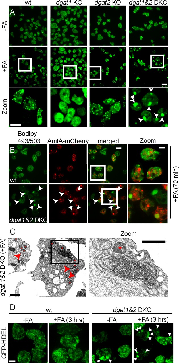
Fig 5. Excess FAs leads to ER-membrane proliferation in dgat1&2 DKO cells.
A. LDs are formed in wild type and dgat2, but not in dgat1 KO cells. Instead of LDs, massive Bodipy-positive structures were observed in the dgat1&2 DKO (arrowheads). FAs were added to the culture medium and a time-lapse movie was recorded with 10 minute frame intervals. Shown are maximum z-projections of 6 sections 1.5 μm apart taken after 180 min. Scale bars, 10 μm. B. The neutral lipid structures in the dgat1&2 DKO (arrowheads) are not of endosomal nature. Wild type or dgat1&2 DKO cells expressing AmtA-mCherry were incubated with FAs and a time-lapse movie with 5 min frame intervals was recorded. Shown is a representative image taken after 70 min. Scale bar, 10 μm; Zoom 5 μm. C. The neutral lipid structures in the dgat1&2 DKO are formed by ER-membranes. Dictyostelium was fed 3 hours with FAs before fixation with glutaraldehyde. Asterisks label mitochondria that have been seen close to the ER-membrane-proliferations. Arrowheads point to long ER-strands. D. GFP-HDEL accumulates in the ER-membrane proliferations in the dgat1&2 DKO. Images of Dictyostelium expressing GFP-HDEL were taken under normal conditions (-FAs) and after 3 hrs incubation with FAs (+FAs). Shown are maximum z-projections. Arrowheads point to ER-membrane proliferations. Scale bar, 5 μm.

