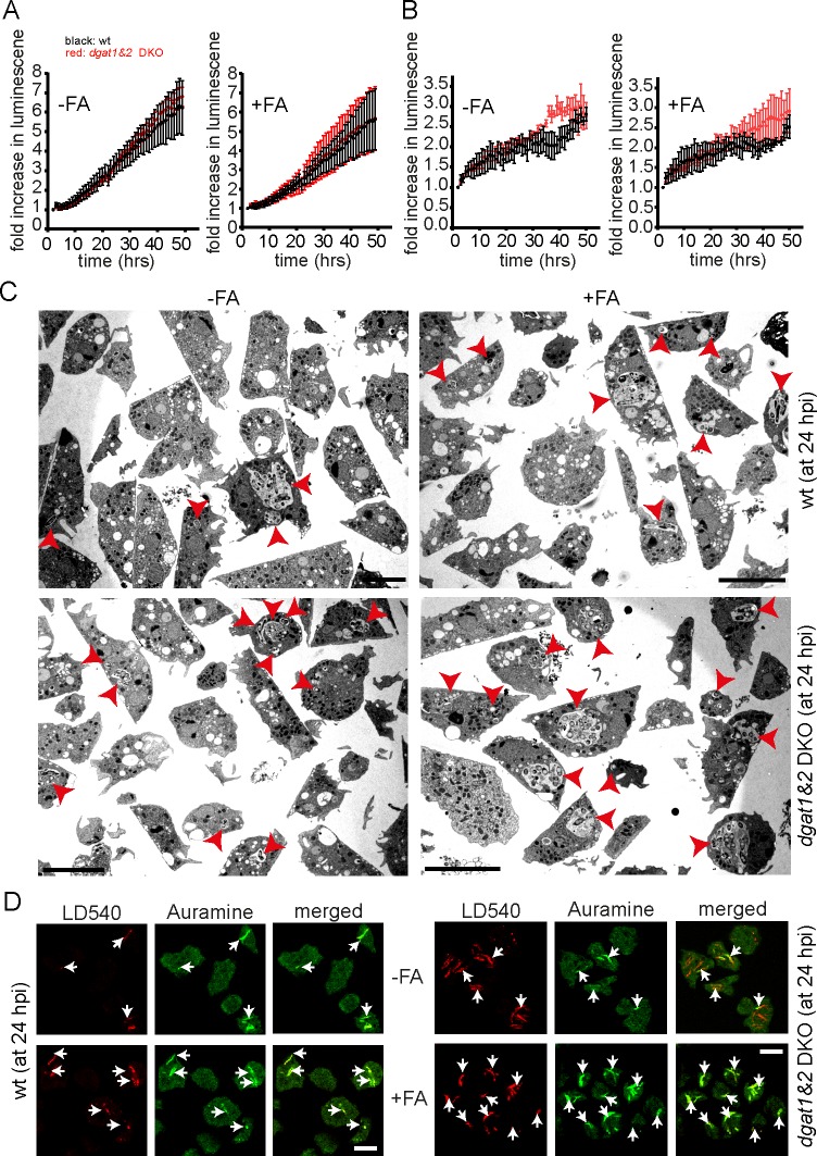
Fig 10. Bacteria are metabolically active and remain acid-fast positive in the dgat1&2 DKO cells.
A. Metabolic activity of M. marinum is unaltered in the dgat1&2 DKO. Wild type and dgat1&2 DKO cells were infected with bacteria expressing bacterial luciferase. Luminescence was recorded every hour with a microplate reader. Shown is the fold increase in luminescence over time. Symbols and error bars indicate the mean and SEM of three independent experiments. A two-way ANOVA test indicated no statistical difference between the curves. B. The number of intracellular bacteria is comparable between wild type and dgat1&2 DKO cells. Dictyostelium cells were infected with mCherry-expressing M. marinum, stained with Bodipy493/503 and plated on 96-well plates. Images were recorded every hour with a high content microscope. After imaging, Dictyostelium cells and bacteria were segmented and analysed. Symbols and error bars indicate the mean and SEM of three independent experiments. A two-way ANOVA test indicated no statistical difference between the curves. C. Dgat1&2 DKO cells harbour more bacteria compared to wild type cells. Wild type and the dgat1&2 DKO cells were infected with unlabelled M. marinum wild type. At 24 hpi, cells were fixed with glutaraldehyde, stained with osmium and further processed for EM. Dictyostelium was fed with FAs prior to infection. Scale bars, 10 μm. D. Bacteria remain acid-fast positive in the dgat1&2 DKO. Cells of wild type Dictyostelium and the dgat1&2 DKO were infected with unlabelled M. marinum. At 24 hpi cells were fixed and subsequently stained with AuramineO and LD540. Cells were fed with FAs prior to infection where indicated. Arrows point to intracellular bacteria. Scale bars, 10 μm.

