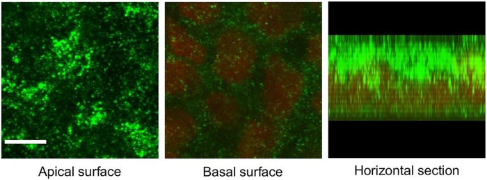
Fig 1. Melanocortin-1 receptor (MC1R) expression in Caco-2 cells.
Cells were grown into confluent monolayers for 21 days on culture inserts and immunostained for MC1R. Nuclei were labeled with propidium iodide. Optical sections prepared by a confocal microscope show MC1R expression levels at the apical and basal surfaces, and a digitized horizontal section of the cells. Green: MC1R labeling; red: cell nuclei. Scale bar: 5 μm.

