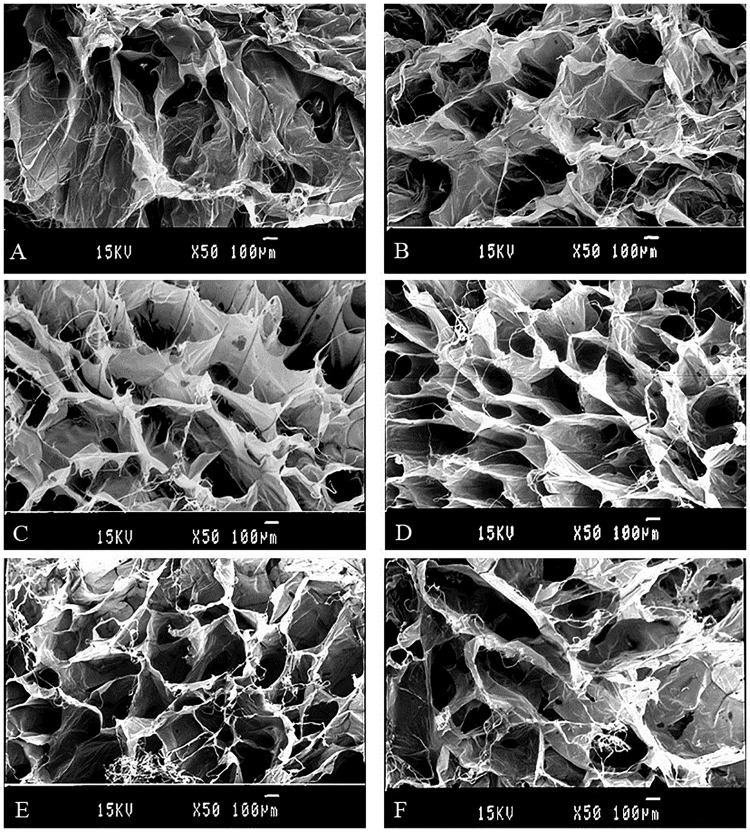
Fig 3. SEM images of cross-linked and uncross-linked lyophilized collagen sponges of different concentration.
A, B: cross-section and surface of 3.3 mg/ml collagen sponges cross-linked by 100mM EDC for 24 h, respectively. C, D: cross-section and surface of 10.0 mg/ml collagen sponges cross-linked by 100mM EDC for 24 h, respectively. E, F: cross-section and surface of 10.0 mg/ml collagen sponges uncross-linked, respectively.

